What is muscle tissue?
Muscle tissue is a complex system of many different kinds of cells. The main component of muscle tissue is the muscle cell. However, the tissue includes nerve, blood, connective, epithelial, stem, and other cells. Because muscles move, they need and use a great deal of energy quickly. Muscle tissue, therefore reconfigures and redesigns cells and organelles in order to provide the energy, protein, and other elements required. Muscle tissue also repositions its cells, especially its subcellular components, mitochondria, and other organelles, to maximize the ability of muscle tissue to contract and to move the body.
Tissue types. There are 3 types of muscle tissue, designated skeletal, cardiac, and smooth. This description is of skeletal muscle cells only.
Muscle cells
A muscle cell is a cell whose shape and composition enable it to become shorter each time it receives an impulse from a nerve ending. A muscle cell has two functions: 1) receive the energy impulse from the nerve cell ending, and 2) react to the energy impulse by shortening itself. The natural state of the muscle cell is flacid; it contracts only if it receives that energy impulse.
Organelles. A muscle cell makes great use of the organelles inside it in that the standard organelles as shown in the generic cell are in many cases completely reconfigured to enable the muscle cell to receive nerve pulses and contract. For example, the nuclei are pressed outwards and protrude from the sarcolema (plasma membrane) as bumps so they don't impede the tightening of the contracting fibers inside the cell interior. Because they appear so different, when the muscle cell was first being discovered and researched, the components of the cell were given their own unique names. It was only later that people realized that these components were the same organelles as in standard cells (see Table 1, below).
In the muscle cell, the endoplasmic (sarcoplasmic) reticulum morphs into flat balloon caverns called cisternae.
| Table 1: Muscle cell component names that parallel standard cell names | ||
|---|---|---|
| In the muscle cell | In a standard cell | Notes |
| Sarcolemma | Plasma membrane | The lipid layer envelope forming the "skin" of the cell. |
| Cytosol (sarcoplasma) | Cytoplasm | Fluid and contents in which float all the organelles. |
| Sarcoplasmic reticulum | Endoplasmic reticulum |
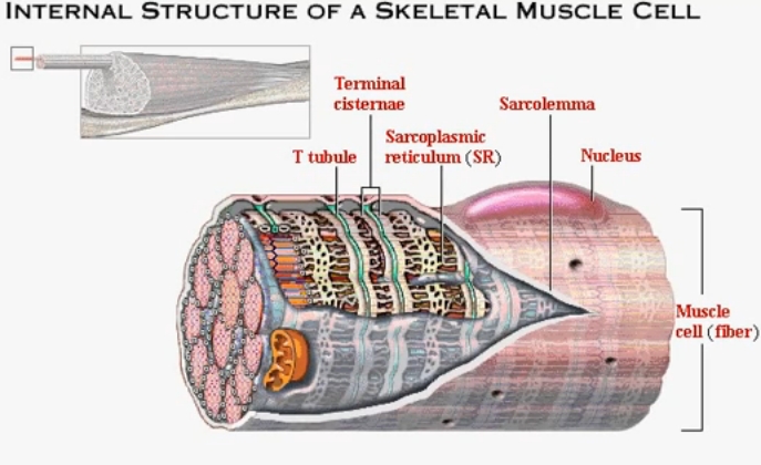
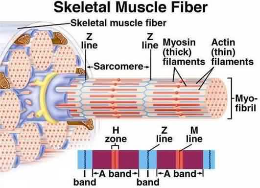
To enable muscle tissue to contract as one unit, as stem cells develop into muscle cells during gestation before birth, what would become individual cells don't get that far. Instead, the internal components of single cells are placed inside one large long thin envelope and become what is known as the muscle fiber. This envelope is usually as long as the specific muscle tissue, stretching from where one end connects into the tendon and bone, to the alternate end, tendon, and bone. The muscle fiber contains the mutiple nuclei of what would have become individual cells, and all the organelles and other components that would have been split among the component cells are now rearranged along the entire fiber for maximum effective contracting ability. Those organelles and tubule/filament structures repeat themselves along the muscle fiber, and each repetition is called a sarcomere.
Receiving the energy impulse
The axons of nerve cells that excite muscle tissue connect to a single location on each muscle fiber, the neuromuscular junction, usually at the center of the length of the fiber. One axon can connect to multiple fibers. The axon terminals pass the action potential into the muscle fiber in the same way as those of other nerve cells pass the potential onto other nerve cells (see Nerves).
Also in the same way as in the nerve cell, the action potential moves rapidly up and down the muscle fiber riding the plasma membrane (sarcolemma) from the central neuromuscular junction to the ends of the fiber.
Each sarcomere has two T (transverse) tubules. These are part of the same lipid layer as the envelope sarcolemma and tube down from that outer membrane into a relatively flat network of connected tubes cross-sectioning the thickness of the muscle fiber.
As the action potential swoops down the muscle fiber, the energy also passes down the walls of the tiny T tubules into the inards of the sarcomere. The T tubules are in close contact with the sarcoplasmic reticulum's central cisternae, and various protein complex gates and channels connect the T tubules to the cisternae on the left and right sides along the tubule. As in nerve fibers, as it passes over, the potential triggers the opening and closing of these various gates/ion channels, in this case, most significantly, opening the calcium ion channel, allowing millions of calcium ions to leave the inside of the cisternae and move into the cytosol. The calcium ions then start a process that enables the micro-filaments inside the sarcomere to pull the ends of the sarcomere together, thus contracting the muscle.
Contracting
The inside of each sarcomere contains thousands of two types of microfilaments, floating in the cytosol and arranged parallel to the muscle fiber. The videos show these better than I could ever describe them. The calcium ions trigger changes in these filaments that make the proteins in one filament (the myosin proteins) reach up and attach themselves to the actin proteins in the adjacent thinner filaments, and pull/slide those filaments towards the center of the sarcomere. This causes the sarcomere (and the whole muscle fiber) to contract.